
Ultrasound is one of the most frequently used procedures in medicine today. Especially in prenatal care, the examination is indispensable, because ultrasound now allows millimeter-precise insights into the development of the unborn.
Table of contents
The Unborn Baby Close-Up In 3D Ultrasound
For a good 30 years, couples have been able to see their unborn baby many weeks before birth and follow its development during pregnancy. As proof of their parenthood, they are then allowed to take a picture with them showing their offspring. For the gynecologist, ultrasound – sonography in medical terms – is an important tool for checking whether the baby is doing well. For this purpose, he sends ultrasound waves into the uterus, which – like all sound waves – are reflected back when they hit something, thus providing an “auditory image” of the sonicated fetus. Ultrasound is therefore also referred to as echography. Bats have shown humans how it works: they use especially emitted ultrasound waves to locate prey and obstacles in pitch darkness. Echo sounders work on the same principle in shipping, where ultrasound was first used by humans.
Does The Baby Feel The Ultrasound?
Ultrasound is not comparable to radiological radiation as in X-rays. The sound waves, with a frequency of 3 to 10 megahertz, are inaudible to humans. Minimal vibrations triggered by the waves do not disturb the unborn child and cannot harm it. Once again, a recent study by the University of Western Australia in Perth gave the all-clear: Even multiple ultrasound examinations during pregnancy have no effect on the baby’s development. The scientists compared the developmental data of children who had five ultrasound examinations during pregnancy with those who had only one sonography. There were no differences in height, neurological development, speech, expression, or social behavior.
What Can Modern Ultrasound Do?
Sound waves are transmitted from the transducer to the uterus via a special gel on the pregnant woman’s abdomen. On their way back, the sensitive transducer picks up the waves again and converts them into electrical impulses. Organs, bones, and tissue appear on the monitor in various shades of gray. The images give the physician a spatial idea of the size and position of the fetus. Today’s devices can even show the vessels of the unborn child, blood flow and velocity as well as the blood exchange between mother and child in different colors in a special color duplex sonography (also: Doppler ultrasound, named after the inventor of this technique). Thanks to a new type of transducer, three-dimensional, plastic images of the uterus are also possible. At the touch of a button, the computer converts the various slice images into a 3D representation. In the special resolution, the three-dimensional images can also capture (as a fourth dimension) a short period of time that reflects the baby’s movements – and provide parents with a short video of their baby upon special request. However, health insurers only pay 50 to 150 euros for a 3D or 4D views in justified cases, such as suspected malformation, growth disorders, or a risk of certain chromosomal disorders.
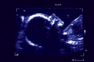
Ultrasound – The Baby In 2D Ultrasound
What Are Ultrasound Scans For?
Three ultrasound examinations, financed by the health insurance fund, are scheduled as part of the usual preventive care appointments: Around the 10th, 20th, and 30th week of pregnancy. Only if the gynecologist notices something, he refers the woman to a special practice or clinic. There, ultrasound experts can examine the unborn child with millimeter precision: In the so-called fine ultrasound, the head and face, spine, extremities, abdomen, gastrointestinal tract, kidneys, and heart of the fetus, in particular, are scanned. The latest generation of equipment is so precise that doctors recently even discovered a clouded eye lens in an unborn baby!
In older and high-risk pregnancies, doctors can now use first-trimester screening, a combination of special ultrasound measurements and a blood sample, to detect possible abnormalities in the unborn child at an early stage (11th to 14th week of pregnancy), such as Down syndrome, genetic malformations or congenital heart defects. This often reduces a theoretically calculated risk of malformation from, for example, 1:100 to 1:3,000. As a result, the need for delicate amniocentesis is much less frequent. Because only if the doctors actually detect something do parents have to decide whether they want further examinations.
Only Experts Are Allowed To Sonicate
All gynecologists who offer ultrasound must obtain a certificate from the Fetal Medicine Foundation and undergo regular training. In order to more clearly emphasize the training and experience of ultrasound practitioners, the “German Society for Ultrasound in Medicine” (DEGUM) has developed three levels of qualification: from basic training (1) to proficiency in fine diagnosis (2) to scientific expertise (3). Pregnant women can calmly ask in the practice what qualification their gynecologist has and whether he can perform detailed malformation diagnostics. After all, how accurate and reliable the ultrasound ultimately depends on the experience of the examiner. The major goal of German ultrasound experts is therefore to be able to offer all women fine diagnostic ultrasound examinations at a specialized center in the future – as is the case in other European countries.
Life Chances Through Ultrasound
The great advantage of early diagnosis is that some of the above-mentioned malformations can now be operated on before birth and give the unborn child a chance to live or survive. If the baby is known to have a disease, the woman can give birth at a special clinic where the newborn can be treated immediately. Unfortunately, even ultrasound still has limitations: Some disorders are not revealed by even a fine diagnostic examination, such as minute heart defects, some kidney damage, or metabolic disorders. In addition, fine ultrasound can also find problems for which medicine does not yet have a solution or doctors cannot intervene before birth. Therefore, some parents prefer not to receive the most accurate examination results and prefer not to know anything. However, it is precise with the help of plastic, three-dimensional images that doctors can often help babies with malformations into the world. With a 3D view of the abdomen, they can better explain the condition to parents and gently prepare them for it.


10 thoughts on “The Baby In Ultrasound – Questions About Ultrasound”